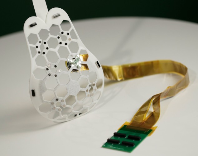
A wearable ultrasound scanner that could help detect breast cancer earlier – and thus improve survival rates – has been developed by a research team headed up at Massachusetts Institute of Technology (MIT).
Breast cancer is the most common cancer to affect women, with around 2.3 million new cases diagnosed worldwide in 2020. When caught in its early stages, the survival rate is nearly 100%, but this drops to 25% among patients whose tumours are detected at a late stage. Regular self-checks and mammograms can help secure positive outcomes. However, in the UK, an estimated 39% of women do not regularly check their breasts for the signs of cancer – which can include shape changes, lumps or swelling.
On the screening front, notes surgical oncologist Tolga Ozmen of the Massachusetts General Hospital, �one of the main obstacles in imaging and early detection is the commute that the women have to make to an imaging centre.†Yet even when women do undergo regular mammograms, breast tumours can still sneak up between screenings. These �interval cancers†make up 20–30% of all breast cancer cases, and their tumours tend to be more aggressive than those found during routine scans.
Galvanized by the death of her aunt – who was diagnosed with late-stage breast cancer at 49, despite regular check-ups – MIT engineer Canan Dagdeviren set about designing an easy-to-use device that could enable more frequent and even at-home screening.
â€Å�My goal is to target the people who are most likely to develop interval cancer,†explains Dagdeviren. â€Å�With more frequent screening, our goal is to increase the survival rate to up to 98 percent.â€Â
To realise this vision, Dagdeviren, Ozmen and colleagues developed a miniaturized ultrasound scanner using a novel piezoelectric material fashioned into a phased array. The scanner fits inside a small tracker that’s inserted into a flexible, 3D-printed patch with a honeycomb-like pattern. The tracker can be moved along a path in the patch to provide imaging from six different positions, together affording coverage of the entire breast.
The whole setup is worn by attaching the patch, via magnets, to a special bra containing openings where the scanner can contact the skin. The researchers note that the device, which they describe in Science Advances, does not require any special expertise to operate.

�We changed the form factor of the ultrasound technology so that it can be used in your home. It’s portable and easy to use, and provides real-time, user-friendly monitoring of breast tissue,†Dagdeviren explains.
To demonstrate their scanner’s potential, the team tested it on a 71-year-old patient with a known history of breast cysts. The wearable enabled the researchers to detect the cysts, which were as small as 3 mm in diameter, the same scale as early-stage tumours. Furthermore, the researchers report, the device achieved a resolution comparable to that of traditional ultrasound systems used in medical imaging centres and could image tissue to a depth of 8 cm.
At present, the scanner has to be connected to a traditional imaging system in order for its data to be visualized. However, the researchers are developing a miniaturized version – about the size of a smartphone – to add to their scanner. Other developments being planned include the integration of artificial intelligence-powered diagnostics and adaptation of the ultrasound technology for use on other parts of the body.
Sheng Xu – a materials scientist from the University of California San Diego who was not involved in the present study, but is also working on conformable ultrasound patch designs – called the device â€Å�impressiveâ€Â. He adds: â€Å�The tracker in the bra could help standardize the ultrasound imaging procedure and minimize operator dependency, which plagues conventional ultrasound technology.â€Â

Wearable coil vest could change the game in breast MRI
�This would be of great benefit to individuals and health services alike,†agrees biophysicist Jeff Bamber of the Institute of Cancer Research, who says that in the UK, pressure on imaging services in hospitals has helped generate waiting lists that are millions of patients long. Bamber notes that the low profile needed for wearables makes it difficult to build ultrasound transducer arrays without compromising on image quality.
He adds: â€Å�This group has incorporated single crystal piezoelectric array technology with advanced doping of the crystal, as used in modern conventional ultrasound probes. The result is good electromechanical coupling, good sensitivity and wide bandwidth, enabling them to achieve relatively good image quality to practically useful imaging depths.â€Â
�With further development to produce images of diagnostic quality, the potential for use in home monitoring would be considerable,†Bamber tells Physics World.

