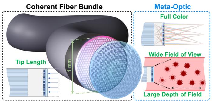
Ultrathin optical elements known as meta-optics can reduce the tip length of endoscopes, which is one of the limiting factors of these medical devices. That’s the latest finding from researchers at the University of Washington, who used an inverse design approach to downsize the tip length by a third. They also demonstrate that the endoscope can capture video in real time over the full visible spectrum, something that has proved difficult with previous approaches.
Endoscopy involves inserting a long, flexible tube (consisting of a camera and a light guide) into the body to obtain images of internal tissues. In existing devices, the tube is tipped with a rigid optical component, the length of which is a fundamental limitation to the device being able to travel through small convoluted ducts such as arteries.
In principle, this problem can be solved by making an endoscope from just a single optical fibre or a bundle of fibres, but the snag here is that some of the light travelling down the fibres is scattered by defects and gets distorted beyond recognition. It cannot therefore be reconstructed to obtain an accurate image. Such devices are also limited to short working distances.
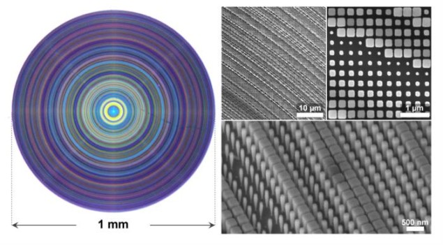
Flat meta-optics provide a promising alternative. These are subwavelength diffractive optical elements comprising nanoscale light scatterer arrays designed to shape an incident wavefront’s phase and amplitude. There is a problem again, however, in that these elements suffer from strong aberrations (or blurring), making large field-of-view (FoV) and full-colour imaging difficult – something that is critical for clinical endoscopy. Indeed, while metalenses typically produce sharp images for a specific wavelength (say green), they strongly blur other colours (red and blue).
Although these difficulties can be solved to some extent by dispersion engineering, the resulting devices suffer from small apertures (around 125 µm, for example), have short working distances (around 200 µm) or require complicated computational post-processing, which makes real-time imaging challenging.
Capturing real-time full-colour images
Researchers led by Johannes Fröch and Arka Majumdar may now have a solution to these challenges with an inverse-designed meta-optics element that they optimized to capture real-time full-colour images with a 1-mm-diameter coherent fibre bundle. Their system allows for a FoV of 22.5°, a depth-of-field (DoF) of more than 30 mm, and a rigid tip that measures just 2.5 mm – that is, 33% smaller than traditional commercial �gradient-index†lens integrated fibre bundle endoscopes. The feat is possible thanks to the shorter focal length and the ultrathin meta-optic.

â€Å�Meta-optics are optical elements that manipulate light in different ways to the lenses that we are used to in everyday life,†explains Fröch. â€Å�Instead of a curved glass surface, meta-optics are composed of small nanostructures that affect how the light is diffracted. This means we can essentially bend it and steer it to specific directions or to have other exotic functionalities.â€Â
Inverse design is an approach in which the structure of the meta-optics is designed based on the required functionality, he adds. �We basically start with the result that we wish and then find the structure that will produce that particular result most closely,†he tells Physics World.
The approach and the fabrication of the meta-optics have to be very accurate and the researchers say they have spent several years developing the right software tools and fabrication conditions to optimize all the steps in the process.
Perfectly suited to endoscopic applications
Achieving full-colour imaging with meta-optics is also extremely challenging since the resolution typically becomes worse as the range of colours is increased. â€Å�Meta-optics often only works for one specific wavelength, but when we started working on this topic, we realized that the resolution of the meta-optic fibre endoscope is ultimately limited by the coherent fibre bundle,†says Fröch. â€Å�We would thus trade off the colour bandwidth with the resolution in just the right way to achieve full-colour imaging comparable to standard lenses for this application.â€Â

Ultrathin photoacoustic imaging probe fits inside a needle
The University of Washington team, reporting its work in eLight, says that meta-optics are perfectly suited to endoscopic applications and could even potentially be exploited to realize much more exotic functionalities, such as hyperspectral imaging or phase-contrast imaging. �They really open up a lot of opportunities and we are now in contact with several other research groups and surgeons to work on many of these possible applications,†reveals Fröch.
Before real-world applications see the light of day, however, he admits that there are still many challenges that need to be overcome. For one, the properties of the meta-optics need to be optimized to achieve an even shorter tip length. â€Å�We also need to figure out a way to better integrate the meta-optics with the endoscope to ensure safe operation,†he says. â€Å�Ultimately, we want to find a solution that allows for a low-cost and scalable integration of meta-optics with the optical fibre so that the devices can be made widely accessible.â€Â

